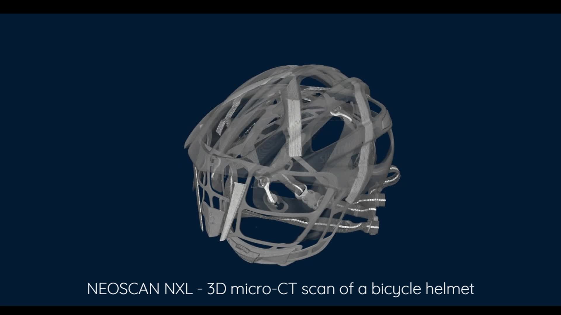
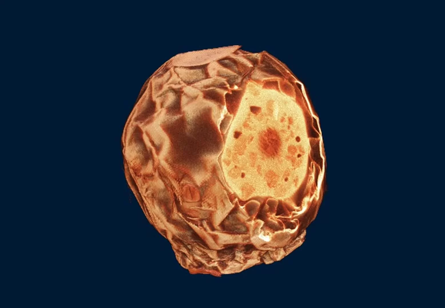
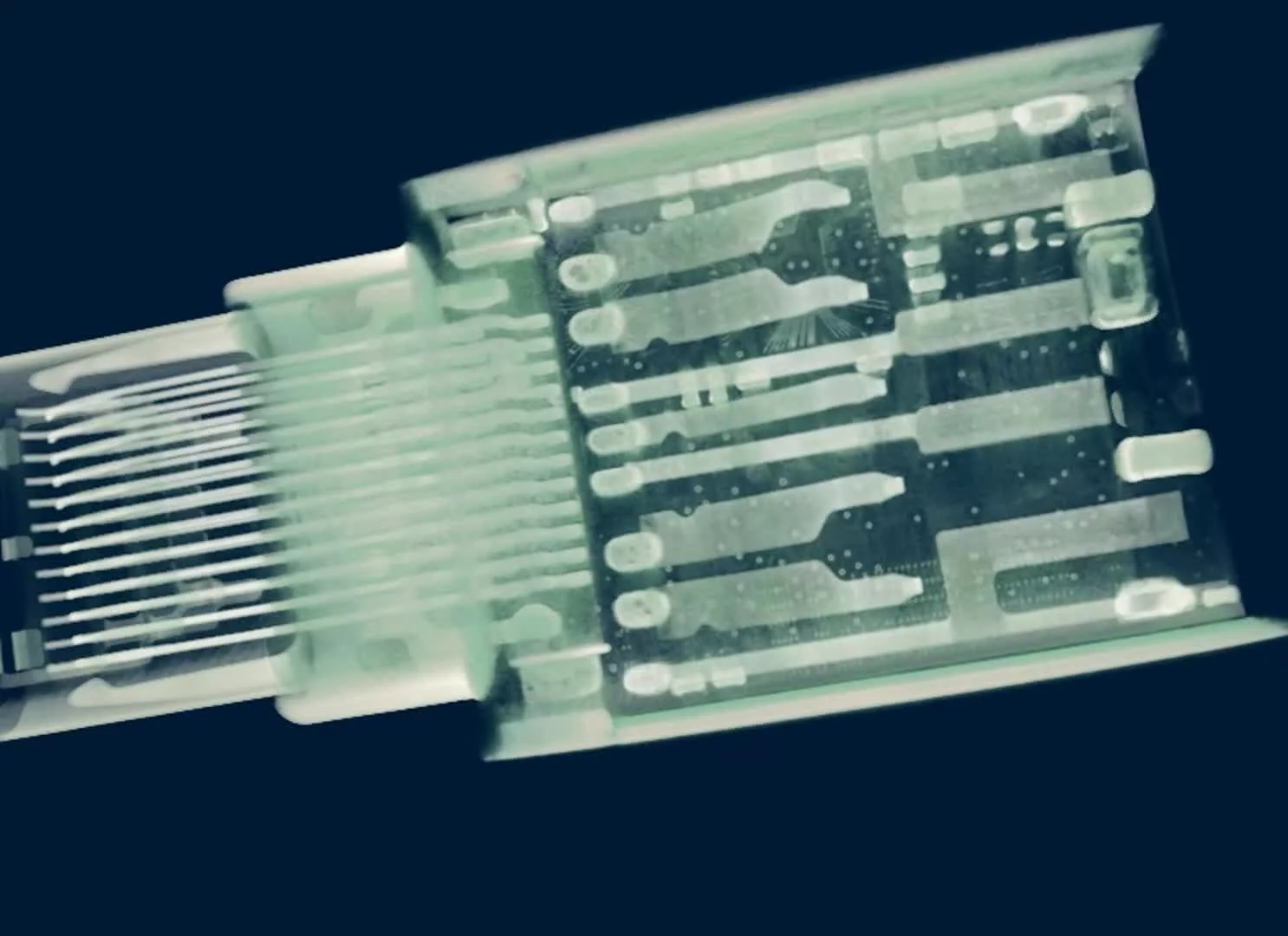
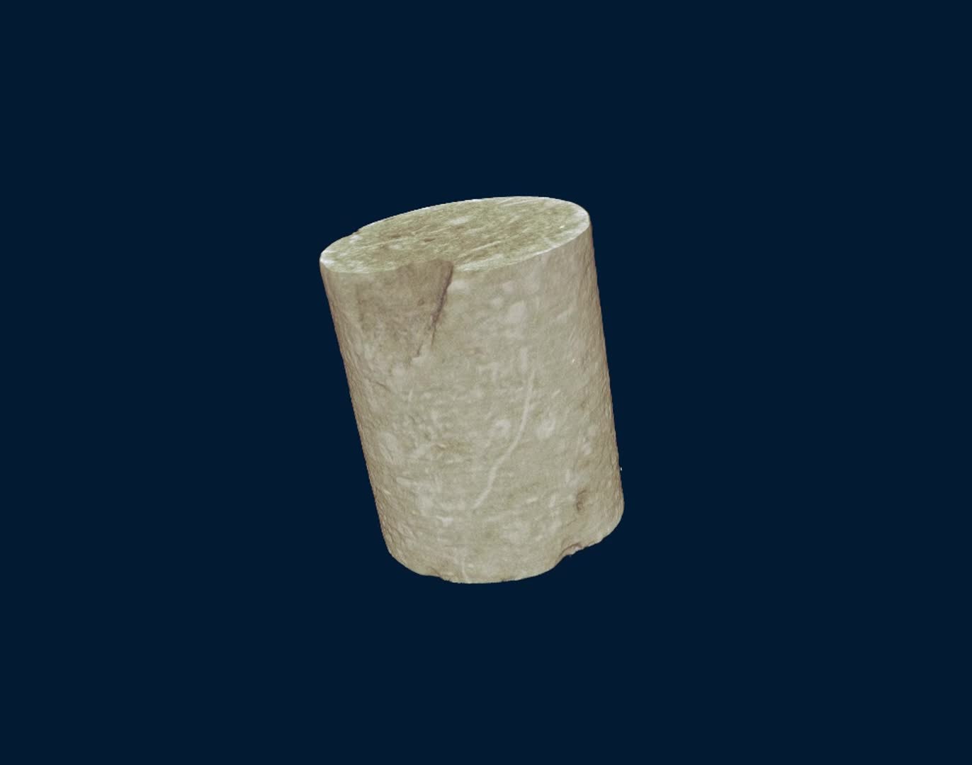
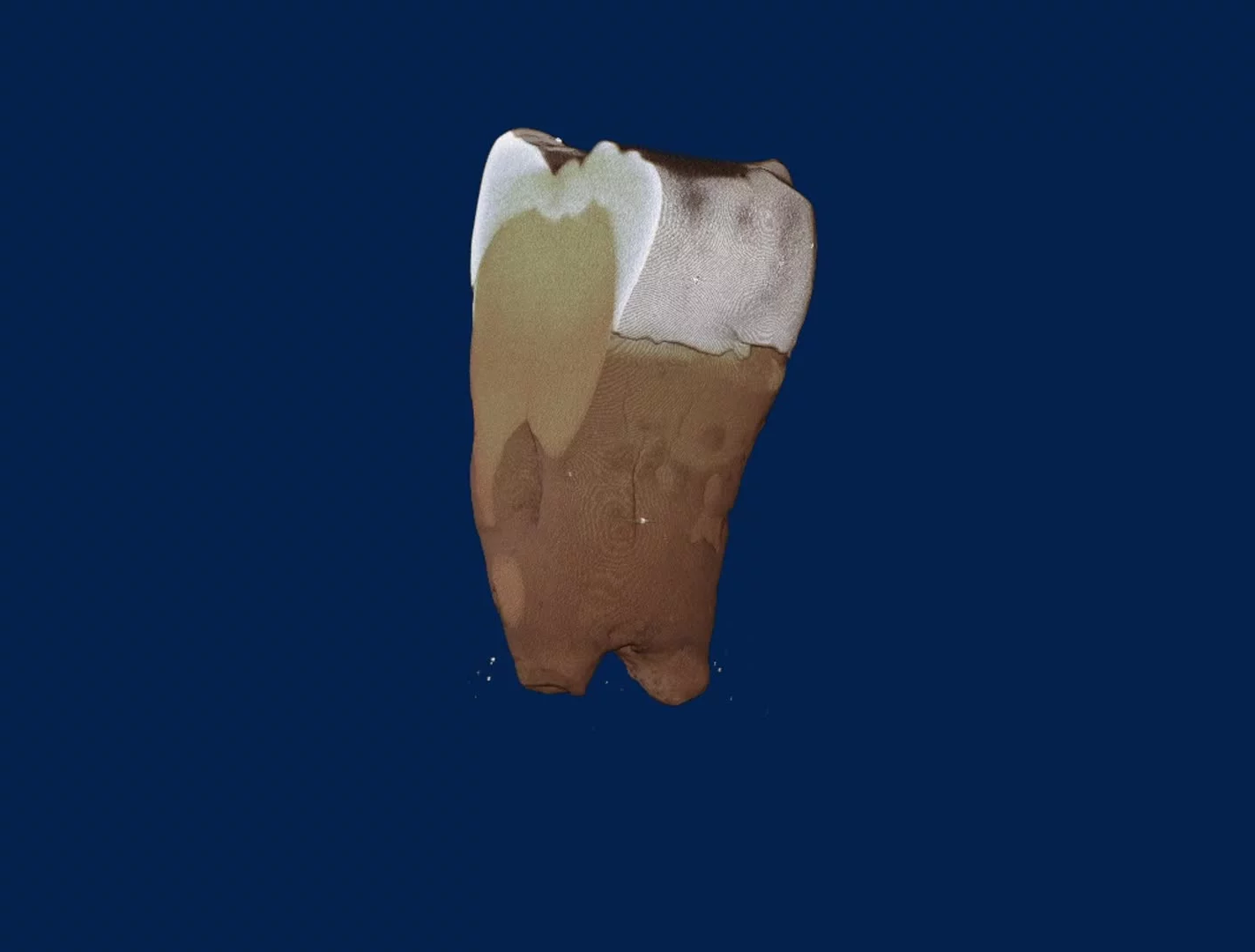
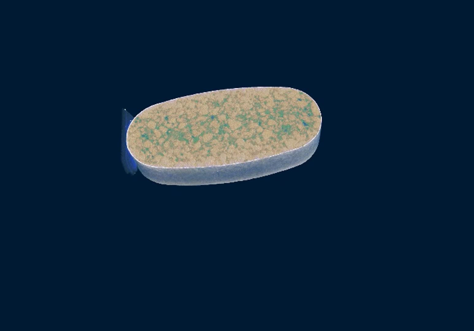
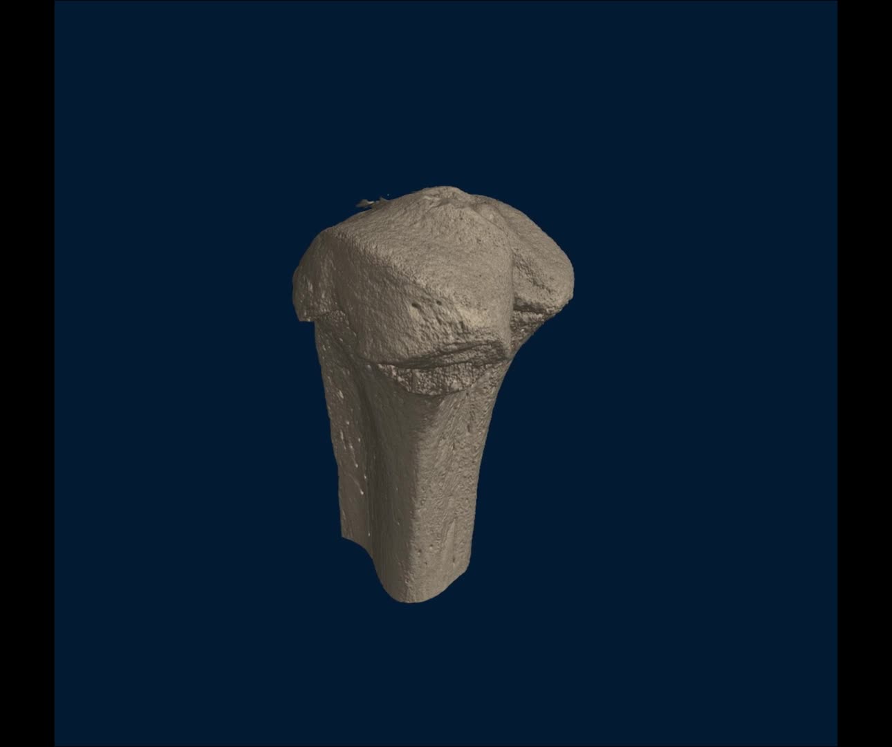
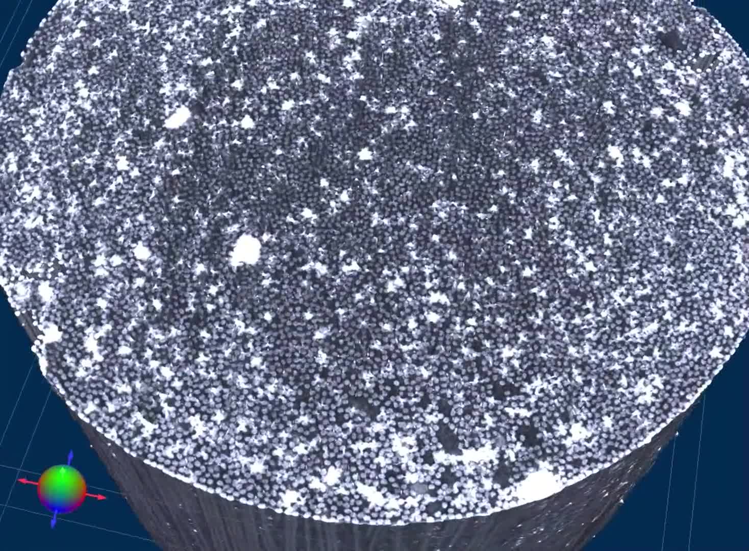
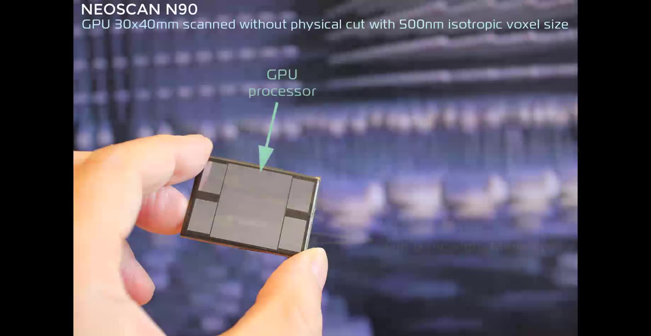
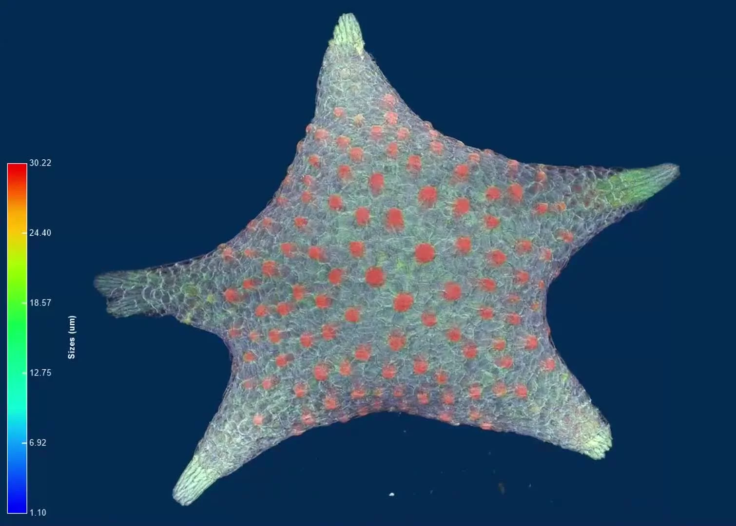
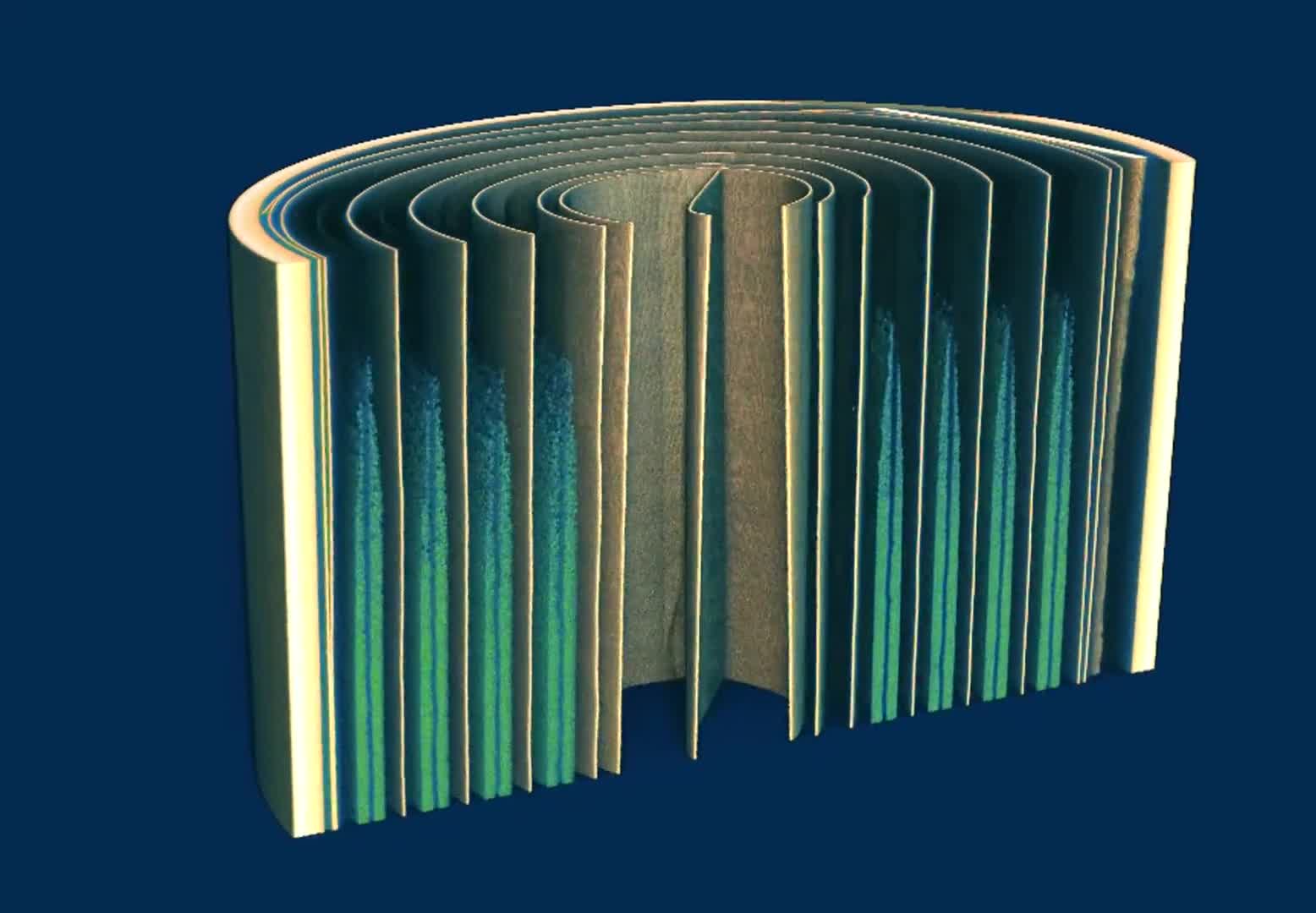
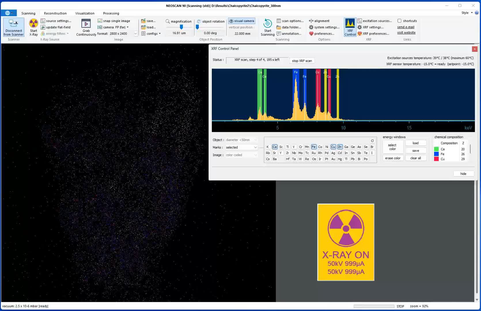
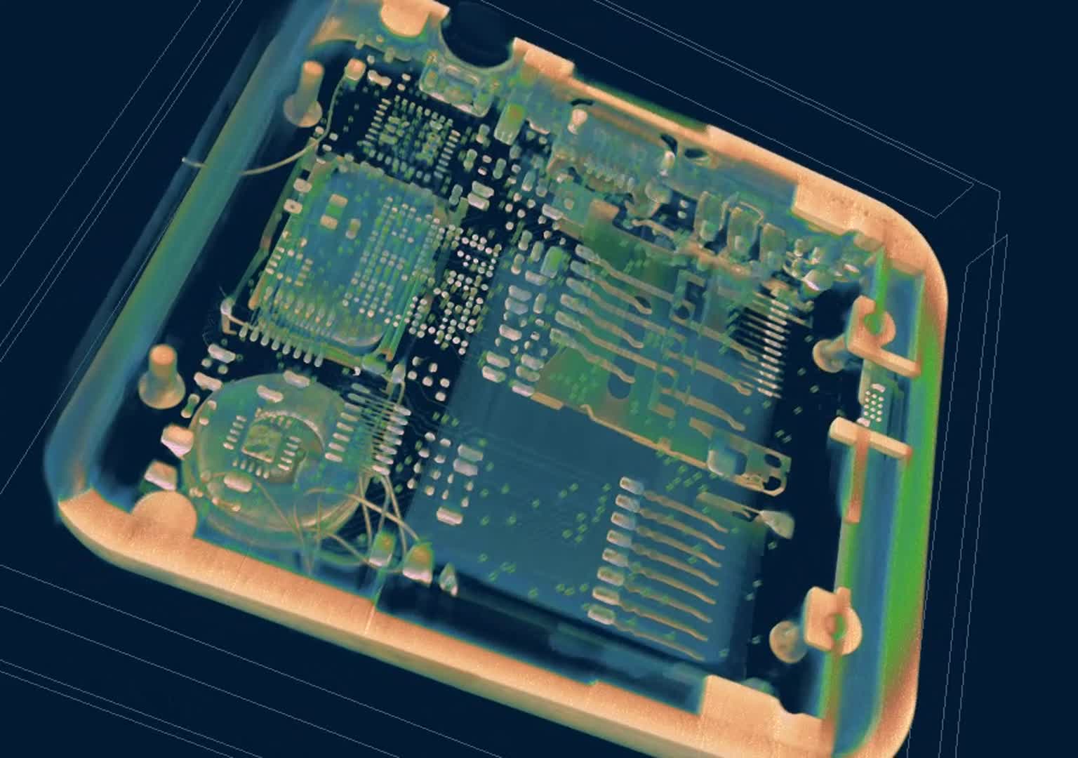
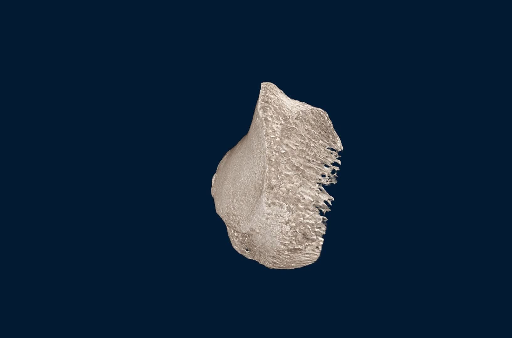
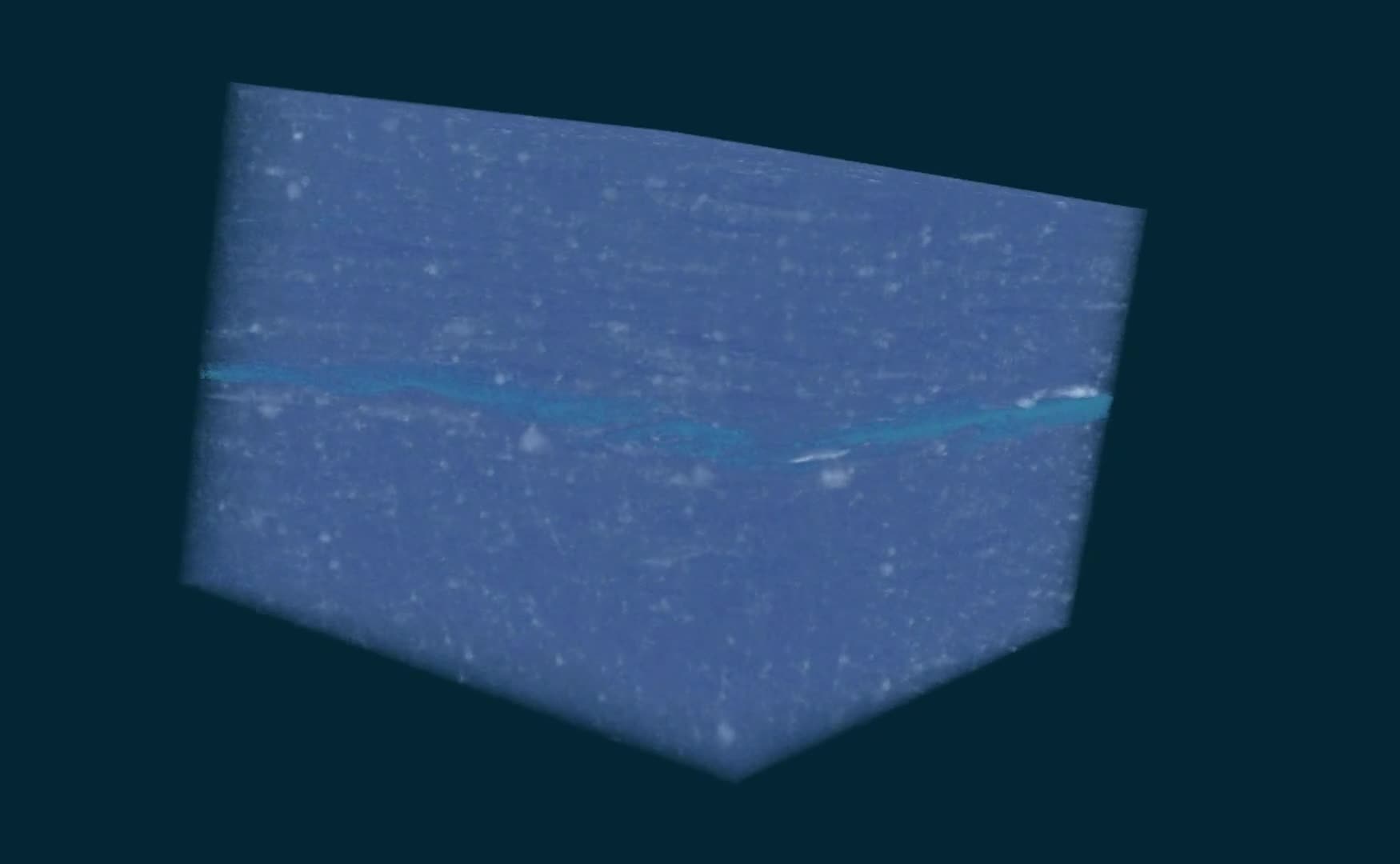
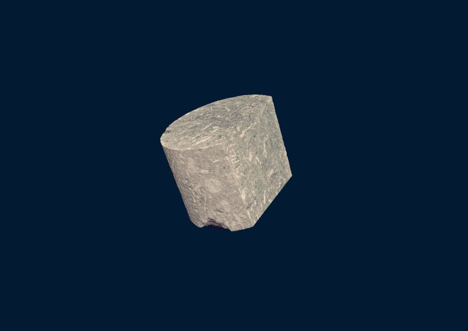
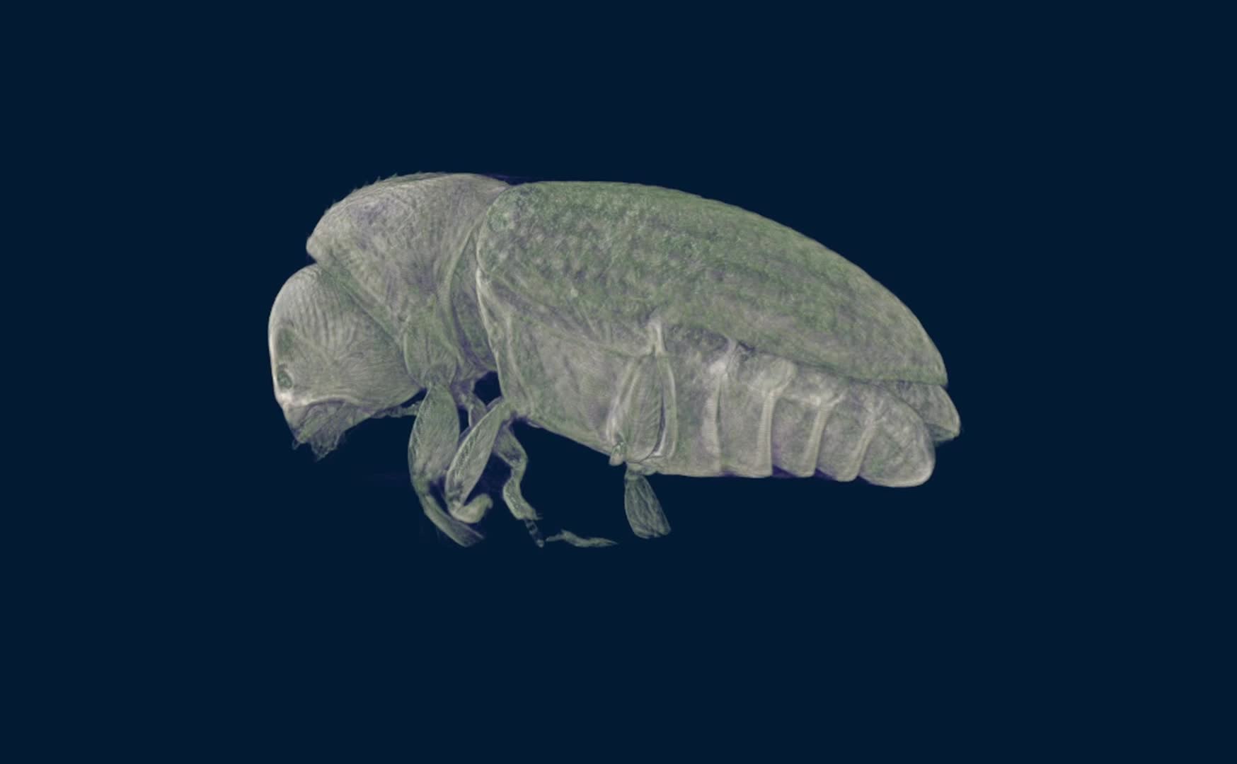
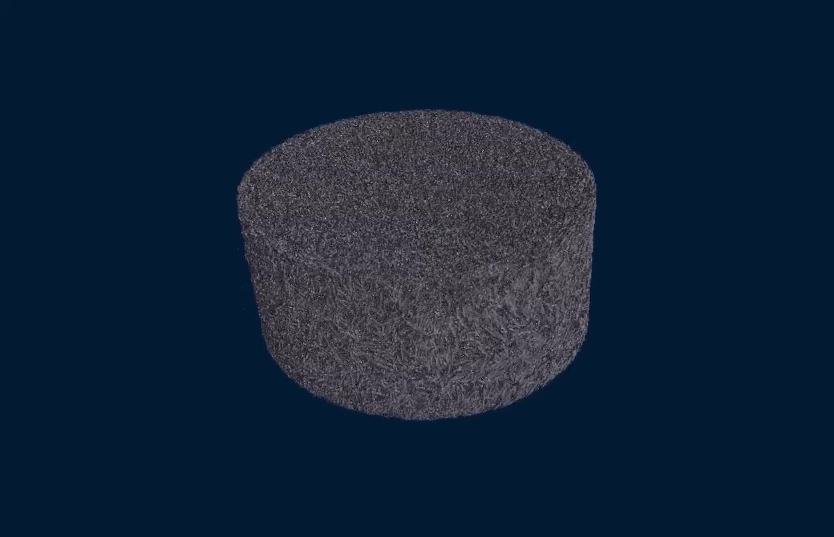
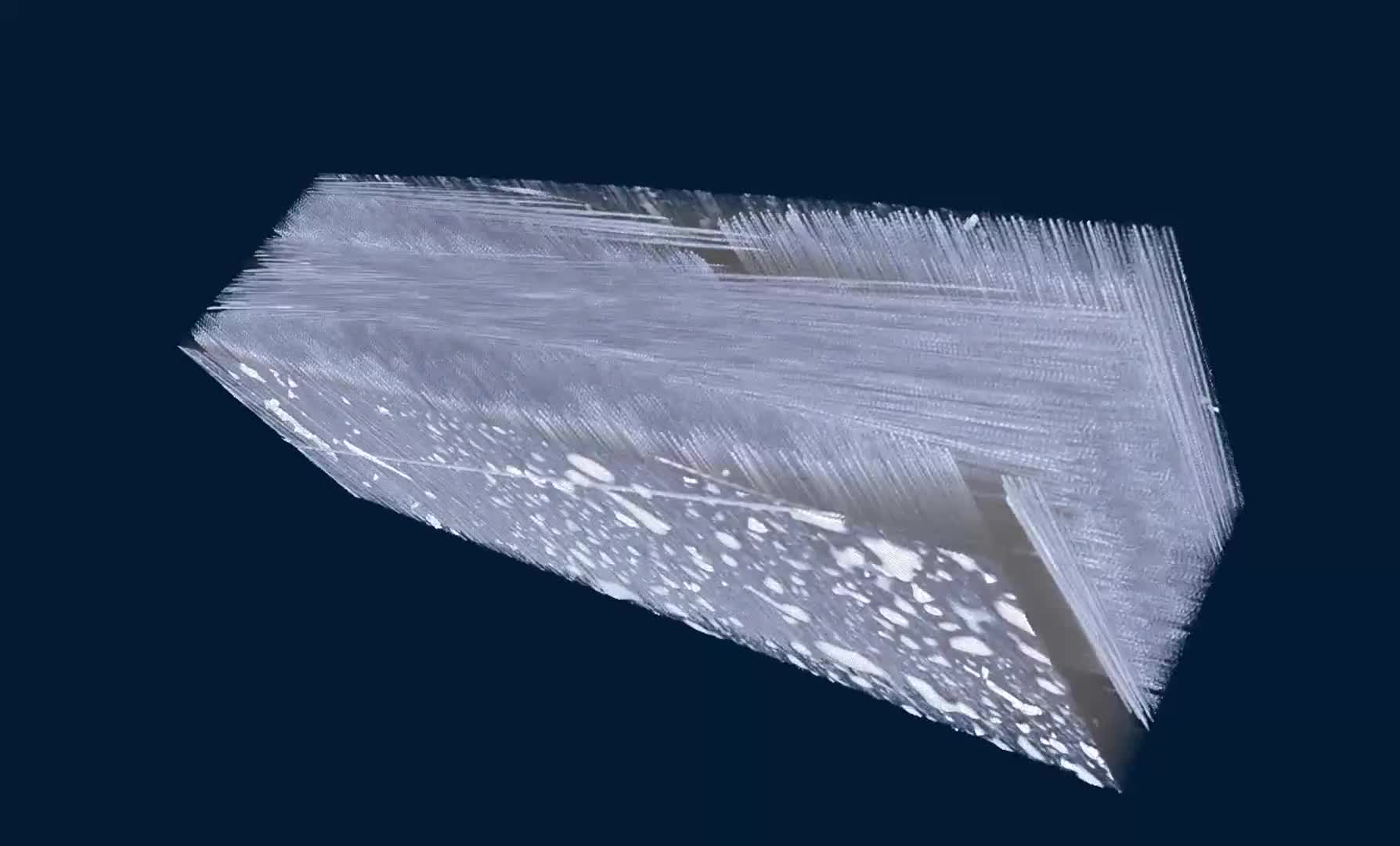
Micro-CT structure of strawberry ice cream in a NeoScan N80 at a pixel size of 7um. The NeoScan Thermal Stage keeps a volume of 3 cm³ of ice cream sufficiently stable at -13°C for high-resolution scanning.
Micro-CT scan of a 32 mm round crunchy biscuit with chocolate and a whole hazelnut inside covered with chocolate and hazelnut crumble. The biscuit is scanned at 19.5 um pixel size.
Non-destructive visualisation of all electronics inside a 128GB USB3 to USB-C flash drive using an N60 desktop microCT scanner.
A carbonate sample scanned at 9.8 um pixel size showing its internal 3D microstructure non-destructively.
A molar tooth scanned at 9.8 um pixel size, showing cracks in dentin and enamel with corroded filing material.
A scan of a pharmaceutical tablet at 9.8um pixel size reveals internal cracks, cracks between tablet and coating and the distribution of active ingredients.
A mouse bone scanned on a Neoscan N70 at high resolution of 3 micron/pixel allows thickness analysis of trabecular bone structures.
3D internal structure of a commercially available type of carbon-fiber reinforced polymer (CFRP). Aside of the large amount of high dense particles in this volume negatively affecting the material's strength, the nano-CT scan at a voxel size of 168 nm provides excellent visualisation of individual fibers, binding material, pores and air inclusions.
Nano-CT scan of advanced GPU with integrated multilayer high-bandwidth memory. Full GPU 40mm in size scanned with 500nm voxels. The scan easily resolves every single one of the TSV’s (Through-Silicon Vias) demonstrating intact connections for communication between memories
Millimetre-sized grain of Star Sand from Okinawa, Japan, the skeletal remains of Foraminifera, scanned with a 550nm voxel size. The nano-CT scan visualizes the 3D morphology and adequately resolves the smallest channels and their connectivity, allowing a 'single micron level' thickness analysis.
Internal structure of a Lithium Ion Battery scanned with 540 nm voxel size
Multilayer ceramic capacitor scanned in a nano-CT N90 at a voxel size of 170 nm, clearly visualizing micron-thickness layers of electrode material separated by ceramic material.
Calcopyrite, scanned with a Neoscan N90. The integrated full-field micro-XRF module allows elemental mapping. The N90 software acquires energy spectra and can produce colour-coded maps of the object’s composition.
A microCT scan at 18 μm pixel size, non-destructively revealing all inner electronics in an entire smartwatch of 50 x 42 x 13 mm.
A microCT scan of a bone at a pixel size of 8 μm, revealing the trabecular structure inside the bone in 3D non-destructively.
MicroCT scan of a shale sample scanned in an N80 at 900nm pixel size, showing the distribution of minerals and the morphology of the breaks between its typical thin laminae.
A carbonate sample scanned at 1.8 μm pixel size, showing its internal 3D microstructure non-destructively.
Coffee borer berry beetle scanned at 980nm, revealing very small internal structures in high detail. Sample courtesy of Prof. J. Alba-Tercedor, University of Granada, Spain
Micro-CT scan of a nylon reinforced with glass fibers. N80 scan with a pixel size of 860nm.