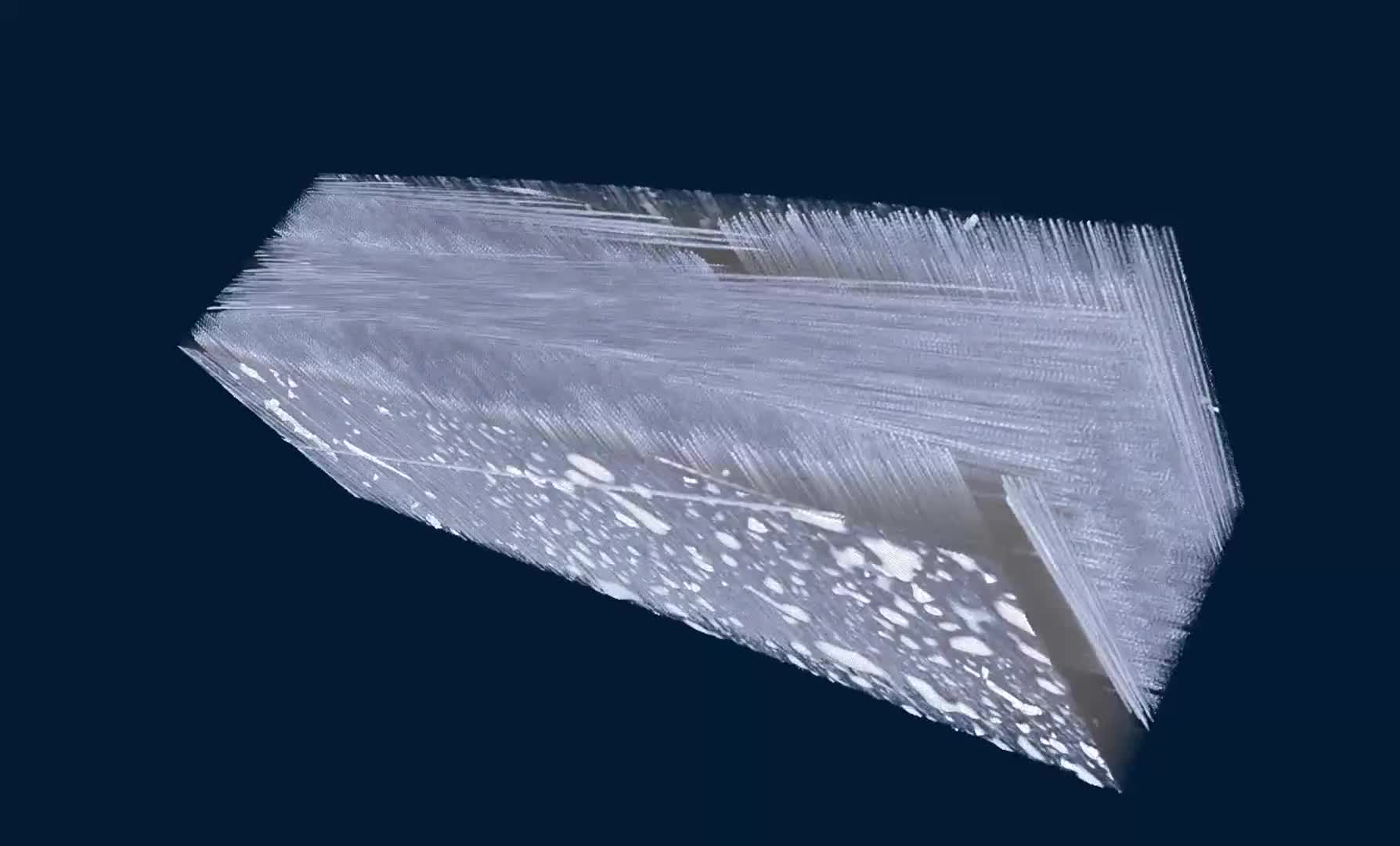
Micro-CT scan of a nylon reinforced with glass fibers. N80 scan with a pixel size of 860nm.
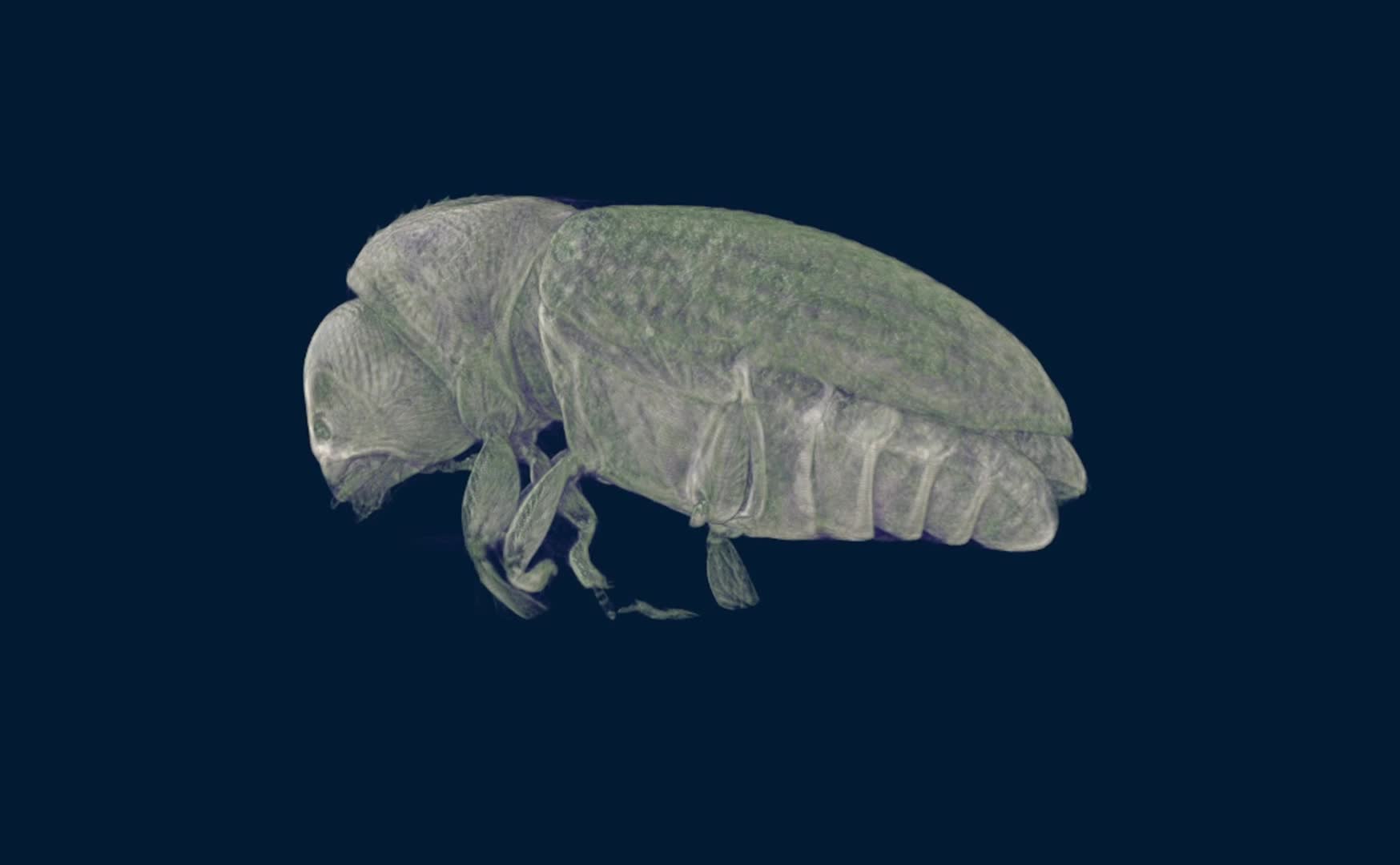
Coffee borer berry beetle scanned at 980nm, revealing very small internal structures in high detail. Sample courtesy of Prof. J. Alba-Tercedor, University of Granada, Spain
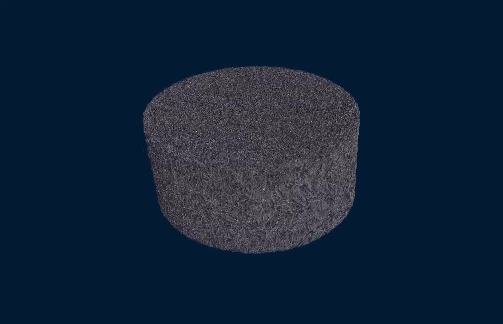
A carbonate sample scanned at 1.8 μm pixel size, showing its internal 3D microstructure non-destructively.
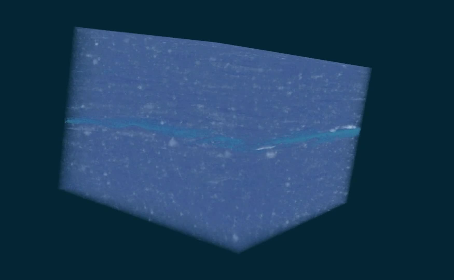
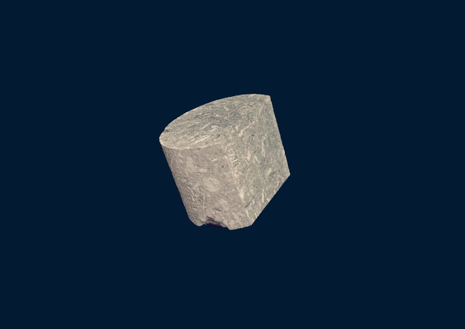
MicroCT scan of a shale sample scanned in an N80 at 900nm pixel size, showing the distribution of minerals and the morphology of the breaks between its typical thin laminae.
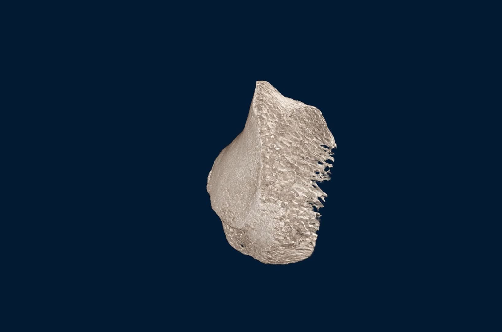
A microCT scan of a bone at a pixel size of 8 μm, revealing the trabecular structure inside the bone in 3D non-destructively.
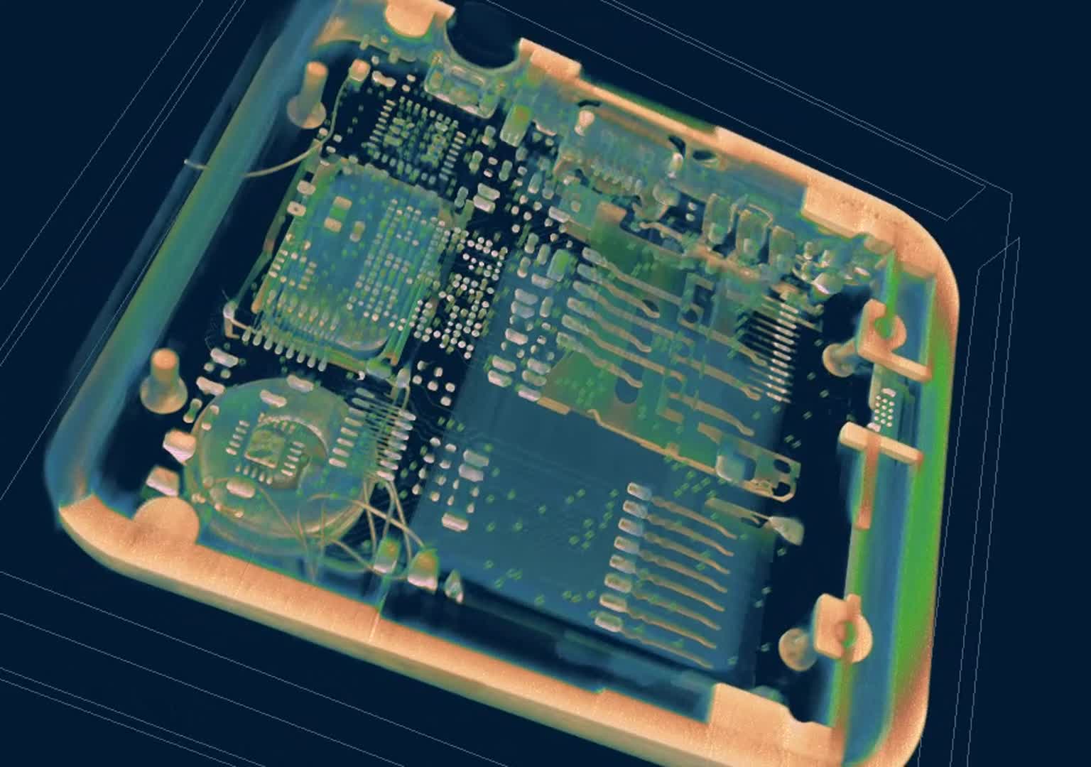
A microCT scan at 18 μm pixel size, non-destructively revealing all inner electronics in an entire smartwatch of 50 x 42 x 13 mm.